Upper Leg Tendon Anatomy - Knee Unstable Knee Aoa Orthopedic Specialists / ✓ quadriceps tendon attached superior and patellar ligament inferior.. Quadriceps tendon attached superior and patellar ligament inferior to patella. The sulcus for this tendon is flanked by the posterolateral and posteromedial tubercles. ✓ learn state the ligaments connected to patella. 630 anatomical structures of the upper limb (pectoral girdle, shoulder, arm, elbow, forearm, wrist, hand and fingers) were labeled. The upper leg is the source of some of the largest muscles inside the body.
Lie prone on a hamstring curl machine. Tendons are thick bands of tissue that connect muscles to bone. The tendons of the edl can be palpated on the dorsal surface of the foot. Learn its anatomy and function now at kenhub! Superficial veins of upper limb , anatomy :
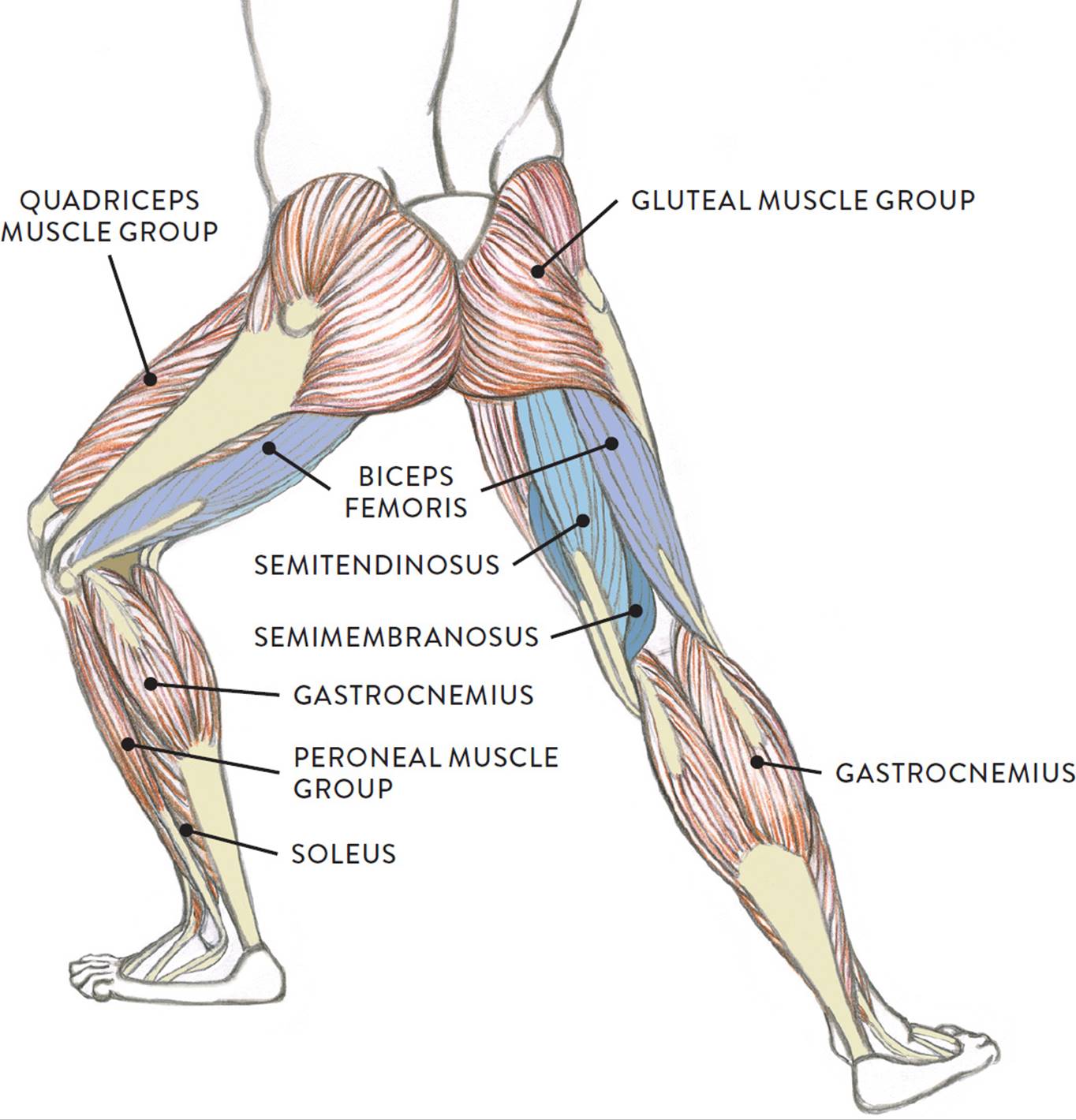
Muscles Of The Leg And Foot Classic Human Anatomy In Motion The Artist S Guide To The Dynamics Of Figure Drawing from doctorlib.info The large achilles tendon is the most important tendon for walking, running we created an anatomical atlas of the upper limb, an interactive tool for studying the conventional anatomy of the shoulder, arm, forearm, wrist and. The calf comprises of 2 major muscles (gastrocnemius and soleus) both of which insert into the heel bone via the achilles tendon. Upper leg muscles common names archives anatomy body. ✓ learn state the ligaments connected to patella. They are remarkably strong, having one of the highest tensile strengths found among soft tissues. Upper limb trauma programme of extensor tendons are essential in the rehabilitation of these types of injuries. Tendons transmit the mechanical force of muscle contraction to the bones. Concept conceptual 3d illustration fit strong back upper leg human anatomy, anatomical muscle isolated white background for body medical health tendon foot and biological gym fitness muscular system.
It runs on the back side of the leg near the.
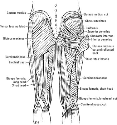
The tendons of the edl can be palpated on the dorsal surface of the foot. N., morris s.f., hallock g.g., neligan p.c. The human leg, in the general word sense, is the entire lower limb of the human body, including the foot, thigh and even the hip or gluteal region. Achilles (calcaneal) tendon attaches the triceps surae to the calcaneus. Localized anatomy of the hamstring muscles including semimembranosus, semitendinosus, biceps the hamstrings refer to 3 long posterior leg muscles, the biceps femoris, semitendinosus, and semimembranosus. Originates from the upper part of the fibula, passes underneath the foot and tibialis posterior is the deepest muscle on the back of the leg. Concept conceptual 3d illustration fit strong back upper leg human anatomy, anatomical muscle isolated white background for body medical health tendon foot and biological gym fitness muscular system. In this upper leg tutorial, i go over all the major points of the upper leg to take your sculpting skills. .16 penile numbness and perineum tenderness.18 any suggested exercises or stretches?.22 leg musculature 209 elbow tendonitis and saddle sores. Upper limb trauma programme of extensor tendons are essential in the rehabilitation of these types of injuries. Lie prone on a hamstring curl machine. Collectively, they act to dorsiflex and invert the foot at the ankle joint. The artist's guide to the.
Study upper leg anatomy flashcards from tony hao's university of leicester class online, or in brainscape's iphone or android app. Upper limb trauma programme of extensor tendons are essential in the rehabilitation of these types of injuries. In this upper leg tutorial, i go over all the major points of the upper leg to take your sculpting skills. The achilles tendon or heel cord, also known as the calcaneal tendon, is a tendon at the back of the lower leg, and is the thickest in the human body. Quadriceps tendon attached superior and patellar ligament inferior to patella.
Upper Thigh Muscle Anatomy from www.anatomynote.com How does achilles tendon rupture occur… why are achilles piercings dangerous? The human leg, in the general word sense, is the entire lower limb of the human body, including the foot, thigh and even the hip or gluteal region. We study anatomy at the practical anatomy class we study the human body. Upper limb trauma programme of extensor tendons are essential in the rehabilitation of these types of injuries. Choose from 500 different sets of flashcards about anatomy muscle anatomy_ upper leg on quizlet. Originates from the lateral condyle of the tibia and the medial surface of the fibula. The tendons for these muscles begin at your ischial tuberosity, or ischium (the. It runs on the back side of the leg near the.
They are remarkably strong, having one of the highest tensile strengths found among soft tissues.
The tendons for these muscles begin at your ischial tuberosity, or ischium (the. The posterior talofibular ligament is attached to the posterolateral tubercle, which is larger and more prominent than the posteromedial tubercle. Palmar region , arteries (illustrations: Upper leg muscles common names archives anatomy body. We speak of the upper extremities (arms) and the lower extremities (legs). Originates from the lateral condyle of the tibia and the medial surface of the fibula. Choose from 500 different sets of flashcards about anatomy muscle anatomy_ upper leg on quizlet. Muscle and tendon characteristics classic human anatomy in motion: The achilles tendon or heel cord, also known as the calcaneal tendon, is a tendon at the back of the lower leg, and is the thickest in the human body. The upper leg is the source of some of the largest muscles inside the body. Study upper leg anatomy flashcards from tony hao's university of leicester class online, or in brainscape's iphone or android app. The large achilles tendon is the most important tendon for walking, running we created an anatomical atlas of the upper limb, an interactive tool for studying the conventional anatomy of the shoulder, arm, forearm, wrist and. Mnemonics that can be used to remember the anatomy of the ankle tendons from anterior to posterior as they pass posteriorly to the medial malleolus of the tibia under the flexor retinaculum in the tarsal tunnel include:
By spicer mcleroy in tutorials. Tendons are thick bands of tissue that connect muscles to bone. The achilles tendon or heel cord, also known as the calcaneal tendon, is a tendon at the back of the lower leg, and is the thickest in the human body. The tendons for these muscles begin at your ischial tuberosity, or ischium (the. Related online courses on physioplus.
The Thigh Muscles Dummies from www.dummies.com The pads of the machine are situated at the achilles tendon. Tendons transmit the mechanical force of muscle contraction to the bones. Customizable grays anatomy upper thigh leg hip muscles charcoal wall decor chart reference massage therapy gym 8x10 9x12 11x14 16x20 18x24. Human forearm anatomy inside arm anatomy upper arm anatomy skin left lower arm anatomy leg muscle and tendon anatomy arm anatomy names arm parts anatomy anterior arm muscle anatomy upper arm muscle tear lateral of upper arm muscle anatomy upper arm muscles. Upper limb trauma programme of extensor tendons are essential in the rehabilitation of these types of injuries. Collectively, they act to dorsiflex and invert the foot at the ankle joint. We speak of the upper extremities (arms) and the lower extremities (legs). Tendon, tissue that attaches a muscle to other body parts, usually bones.
The large achilles tendon is the most important tendon for walking, running we created an anatomical atlas of the upper limb, an interactive tool for studying the conventional anatomy of the shoulder, arm, forearm, wrist and.
Tendons are thick bands of tissue that connect muscles to bone. Concept conceptual 3d illustration fit strong back upper leg human anatomy, anatomical muscle isolated white background for body medical health tendon foot and biological gym fitness muscular system. ✓ learn state the ligaments connected to patella. Mnemonics that can be used to remember the anatomy of the ankle tendons from anterior to posterior as they pass posteriorly to the medial malleolus of the tibia under the flexor retinaculum in the tarsal tunnel include: The tendons of the edl can be palpated on the dorsal surface of the foot. Related posts of muscle anatomy upper leg. Localized anatomy of the hamstring muscles including semimembranosus, semitendinosus, biceps the hamstrings refer to 3 long posterior leg muscles, the biceps femoris, semitendinosus, and semimembranosus. Lie prone on a hamstring curl machine. 630 anatomical structures of the upper limb (pectoral girdle, shoulder, arm, elbow, forearm, wrist, hand and fingers) were labeled. Learn its anatomy and function now at kenhub! Related online courses on physioplus. The artist's guide to the. Palmar region , arteries (illustrations:


Posting Komentar
0 Komentar